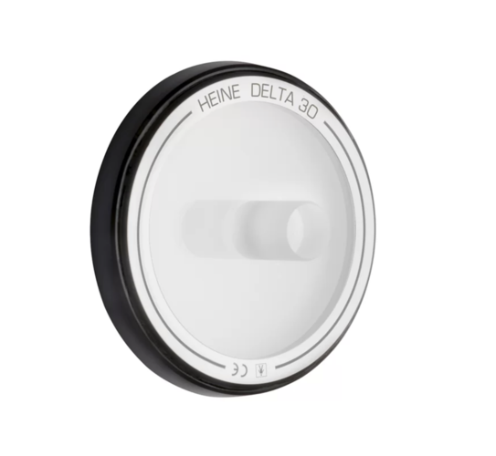
Small Contact Plate for the Heine Delta 30
| katalogové číslo: | 101389.0 |
| EAN: | 4053755197132 |
| výrobce: | HEINE |
| cena bez DPH: | 4 780,992 CZK |
Small Contact Plate for the HEINE DELTA 30
8mm kontaktní kotouč
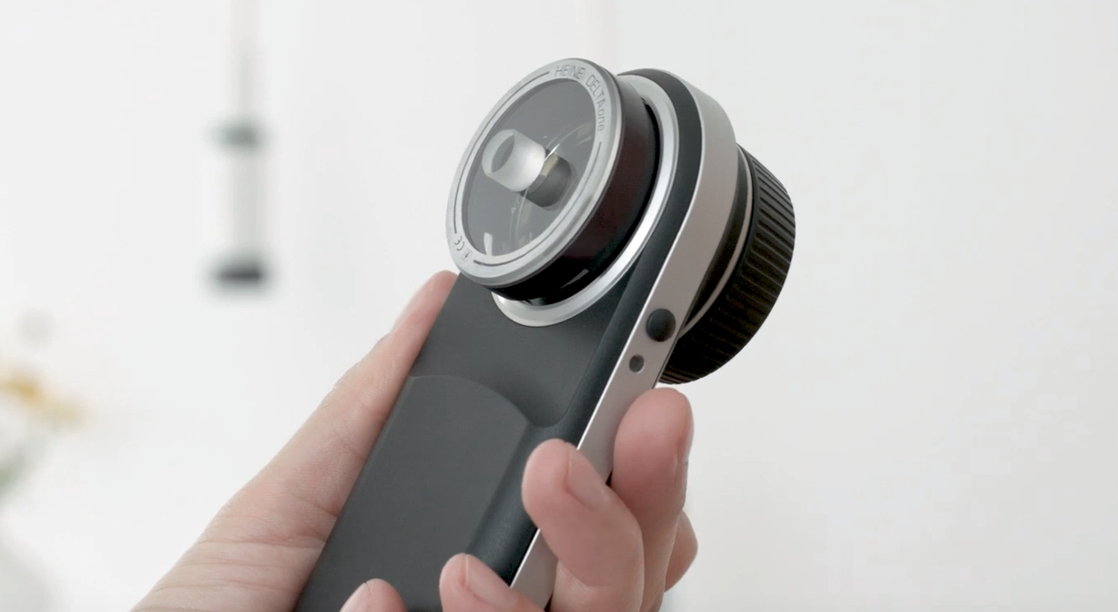
Pro použití s dermatoskopem DELTA 30
Lze použít s imerzní kapalinou (nepolarizovaný režim)
Magnetické uchycení
Snadná a rychlá výměna kontaktní desky
Ideální pro těžko dostupná místa na těle
Např. mezi prsty na rukou a nohou nebo nosní dírky
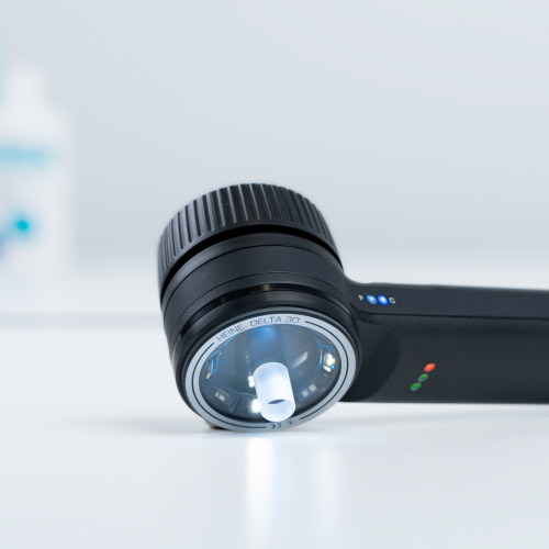
A SMALL CONTACT PLATE MAKES DIAGNOSING EASIER
Dermatoscopy of lesions between fingers and toes
The dermatoscope is considered to be the most important instrument in the dermatologist’s auxiliary equipment toolkit. It has become a vital part of the process of reliable assessments of unpigmented and pigmented lesions, such as naevus cell naevi and melanomas in the context of skin cancer screening. But the dermatoscope is also an important instrument when it comes to detecting other lesions such as plantar warts, hypertrophic sebaceous glands or itch mite infestations. However, reaching prominent places with a dermatoscope is not always straight forward. Often, the clinician cannot get close enough with the dermatoscope in contact mode to sufficiently examine some areas of the skin like for example in interdigital areas, in the ear, in the corners of the eyes or the nasal wings.
In situations such as this it is advisable to use a smaller contact plate like the one available for the HEINE DELTAone or DELTA 30 dermatoscopes. The narrow optical attachment sits perfectly in concave-shaped body parts, therefore enabling dermatoscopy there too.
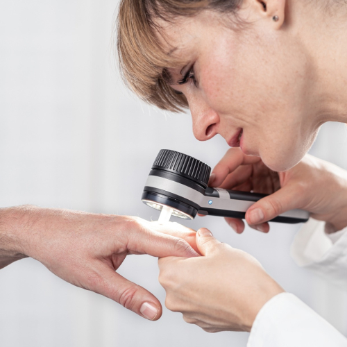
It is impractical in particular to examine lesions between the toes using a normal dermatoscope in contact mode. Pigmented lesions such as naevus cell naevi (which require clarification) are frequently found here too. A dermatoscope with a small contact plate is ideal for easily distinguishing Verrucae vulgares (plantar warts) which also develop on the sides of the toes from foreign bodies or calluses (corns).
Dermatoscopic facial examinations
A smaller dermatoscopic plate is the perfect partner for examining exposed parts of the face – the nose wings, inside and behind the auricle, as well as in the visible external ear canal – Dermatologists often observe white skin cancer (such as basal cell carcinomas) or precancerous white skin cancer (including actinic keratoses) on sun-damaged skin in these areas.
Purely cosmetic lesions such as hypertrophic sebaceous glands, xanthelasma or milia often occur on the forehead, in the middle of the face, in the inner corners of the eyes, on the eyelids or in the nose area. A small, suitable dermatoscope attachment is helpful when dealing with these localised areas which can be difficult to examine with a standard dermatoscope, particularly in light of the fact that these lesions can clinically resemble a basal cell carcinoma, a white skin cancer that is frequently found at the nasal wings or corners of the eyes.
Scabies: on the trail of the itch mite
Itchy red papules, blisters and scratch marks are all indications of an itch mite infection. Itch mites – tiny spider-like creatures that dig corridors 0.5 to 2 millimetres wide under the skin – like to nest in the spaces between the fingers or in the navel. In this situation, a more acccurate diagnosis can be made with a small contact plate used in direct contact with the skin and with immersion fluid. This then allows a dermatologist to reliably distinguish an itch mite infestation from other disease patterns such as eczema in the context of neurodermatitis.
")
")


Dnes představujeme: Elektrický vyplachovač zevního zvukovodu Propulse
Elektrický vyplachovač zevního zvukovodu Propulse Správná péče o ušní hygienu je klíčová pro prevenci a léčbu různých ušních onemocnění. Elektrický vyplachovač zevního zvukovodu Propulse představuje moderní řešení pro efektivní a bezpečné čištění uší, vhodné pro použití v lékařských ordinacích i domácím prostředí. Klíčové vlastnosti vyplachovače Propulse: • Moderní technologie: Elektrický systém zajišťuje ...
Číst celé")
Dnes představujeme: Stetoskop MDF® 777 MD One Stainless Steel Premium Dual Head (MDF 17 Burgundská)
Stetoskop MDF® 777 MD One Stainless Steel Premium Dual Head (MDF 17 Burgundská) Stetoskop je nezbytným nástrojem pro každého zdravotnického profesionála, který umožňuje přesné poslechové vyšetření pacientů. Dnes vám představujeme model MDF® 777 MD One Stainless Steel Premium Dual Head v elegantní burgundské barvě, který kombinuje špičkovou kvalitu, vynikající akustické vlastnosti a atraktivní design. Klíčové vlastnosti stetoskopu MDF® 777 MD One: • Oboustranná hlavice: Umožňuje vyšetření ...
Číst celé
Dnes představujeme: Dětský inhalátor VITAMMY GATTINO A1503 Pink – roztomilý spojenec v boji s respiračními potížemi
VITAMMY GATTINO A1503 Pink Dětský inhalátor VITAMMY GATTINO A1503 Pink je navržen tak, aby zpříjemnil léčbu respiračních onemocnění nejmenším pacientům. Jeho atraktivní design ve tvaru kotěte a přívětivá růžová barva pomáhají dětem překonat strach z inhalace a činí z léčebného procesu hru. Klíčové vlastnosti inhalátoru VITAMMY GATTINO A1503 Pink: • Přátelský design: Tvar kotěte a saténové pouzdro příjemné na dotek zvyšují důvěru dětí při inhalaci. • Univerzální ...
Číst celé
Dnes představujeme: Woodova lampa IDEALDERM
lampa IDEALDERM – světlo, které odhalí víc, než vidí oko Pokud se věnujete péči o pleť nebo krásu profesionálně, nebo jen chcete mít důkladnější přehled o stavu své pokožky, bude vás zajímat Woodova lampa IDEALDERM. Tento šikovný diagnostický přístroj využívá UV záření pro zobrazení různých typů kožních změn, které nejsou pouhým okem vždy viditelné. S její pomocí snadno odhalíte: • typ pleti a její potřeby (mastná, suchá, dehydratovaná) • přítomnost ...
Číst celé


Medicalis - Mgr. Petr Oravski
U lesa 772,
73401 Karviná 4,
Czech Republic